The Ottawa Hospital performs awake brain surgeries approximately three to four times a week. Recent advancements in our brain mapping techniques have allowed us to achieve even more success with this procedure. Today, our team of experts train other surgical teams from across Canada to perform awake brain surgeries using advanced mapping techniques and anesthesia protocols.
What is awake brain surgery?
Awake brain surgery is a highly specialized surgical procedure utilized when operating on the brain with the patient awake and alert, so speech, cognition, movement and sensation can be tested and monitored during surgery. The goal of this technique is to identify and preserve these critical brain functions as their locations can be quite variable between patients.
Awake brain surgery is usually undertaken to remove tumours within the brain including primary brain tumours ranging from astrocytomas and oligodendrogliomas to glioblastomas. Often, these types of tumours have poorly defined borders and can spread into the adjacent brain, making it very difficult to remove them entirely without damaging critical areas. Awake surgery allows the surgeon to identify and avoid these regions while removing the tumour.
With the patient awake and able to respond to specialized tests, the neurosurgeon can identify exactly which areas of the brain are involved with speech, cognition, movement, and other functions. The patient might be asked to perform tasks such as counting, reading, naming pictures, and moving different parts of the body.
“I kept talking, laughing, and singing Disney songs, like ‘Hakuna Matata.’ I was telling them how I was going to go to Disney World when it was over. Five hours seemed like just one.”
— Kimberly Mountain, who received awake brain surgery at The Ottawa Hospital
If the patient is awake, won’t they feel it?
Awake brain surgery requires an entire team of skilled individuals including a neurosurgeon and a specialized neuroanesthetist who work together in conjunction with the nursing team to ensure the comfort and safety of the patient. The neuroanesthetist ensures that the patient is able to interact and participate in testing in a relaxed manner during surgery. Patients are often sedated at the beginning and end of the procedure but are awake during the testing which allows the brain around the tumour to be “mapped”. Testing is continued throughout the tumour removal to ensure that the tumour can be removed as safely as possible.
What is brain mapping?
Brain mapping utilizes various testing protocols for the specific functions of the brain that can be deduced from the patient’s preoperative MRI scan and clinical examinations. Testing specifically involves the application of a small electric current to the surface of the brain or to its deep connection which temporarily can interrupt normal function for one or two seconds. When the current is turned off the function immediately returns to normal. The presence or absence of dysfunction during testing allows the creation of a local brain map. The neurosurgeon uses this map as a guide, along with 3D computer images of the brain, to safely remove the tumour during surgery.
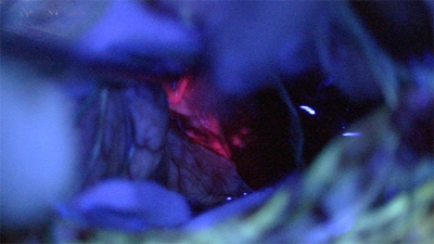
Fluorescence-Guided Surgery
Neurosurgeons at The Ottawa Hospital can now incorporate 5-ALA Fluorescence-Guided Surgery for surgical resection of malignant gliomas using a new, donor-funded, fluorescence-guided microscope. This technique causes the tumour to “glow” or fluoresce under a special wavelength of light generated by the microscope. Since the normal brain tissue does not glow, surgeons can differentiate the tumour margins much more precisely, often identifying and removing tumour that would not have been seen with a traditional operating microscope. This allows for maximal resection of the tumour which leads to improved survival and outcomes.