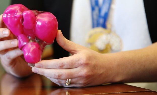
The uterus fits in the palm of Dr. Sony Singh’s hand. The large pink lumps inside the clear, plastic 3D-printed model are fibroids, or tumours, and there are more than 50 of them. To ensure his patient could carry a child in the future, Dr. Singh had to do something that had never been done before.

Maureen had suffered for years with abdominal pain. Over the past six years, she was told by five doctors that she had so many fibroids in her uterus, her only option was to have a hysterectomy – complete removal of her womb. She refused this option.

“I will die with my womb. Nobody will touch it,” said Maureen (who did not want her last name used).

She was referred to the Shirley E. Greenberg Women’s Health Centre at The Ottawa Hospital, where she saw the Minimally Invasive Gynecology team of doctors and nurses. Dr. Singh, a surgeon and the Elaine Jolly Research Chair in Surgical Gynecology, told Maureen he could remove all the fibroids, and she would not need a hysterectomy.

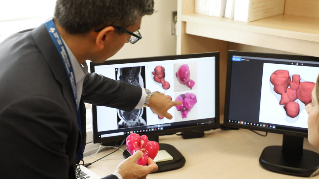
Dr. Sony Singh uses 3D printed model for complex surgery.
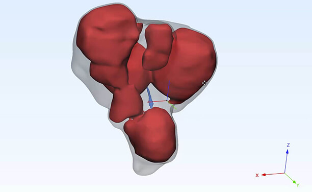
Holding the 3D printed model of Maureen’s uterus, Dr. Sony Singh examined the MRIs and 3D renderings – the images that appear on the operating room screens that doctors can move to get a 3D view of the surgical area.

“Maureen had close to 50 fibroids and we wanted to make sure her uterus was able to carry a baby in the future and function normally,” said Dr. Singh. “But we needed help to plan the complicated surgery to remove them.”

Dr. Teresa Flaxman, Research Associate at The Ottawa Hospital, said it was difficult to see tumours in the patient’s uterus on an MRI. So, she contacted the hospital’s 3D Printing Lab. She had heard how 3D-printed models were helping orthopaedic surgeons see exactly what they were operating on, so they could better plan the surgery.

In 2016, thanks to a donor’s generosity, The Ottawa Hospital acquired a medical 3D printer that uses acrylics and plastics to create exact replicas of patients’ bones and organs from a CT scan or MRI. With the opening of the 3D Printing Lab in February 2017, the hospital became the first in Canada to have an integrated medical 3D-printing program for pre-surgical planning and education.

Dr. Adnan Sheikh, Director of The Ottawa Hospital’s 3D Printing Program, said the Department of Orthopaedics is one of the main users of the lab, which prints models for orthopaedic oncology surgeons to plan operations in advance, reducing surgery times and costs.

“3D printing is revolutionizing the way we look at anatomy,” said Orthopaedic Surgeon and Oncologist Dr. Joel Werier, who has used 3D-printed models of his patients’ hips and bones since the lab opened. “It adds another perspective to how we view tumours, how we plan our surgery techniques, and our ability to offer precision surgery.”

Bones are relatively easy to create from CT scans and MRIs, said Dr. Flaxman. However, soft tissues, such as uterine tissue, is harder to identify, and a model hadn’t been made of one before.

“We’re going to be one of the first hospitals internationally to study how we can provide this improved care by using 3D-printed models in planning surgery for women’s health.”

Dr. Flaxman and other researchers from the Women’s Health Centre worked with Waleed Althobaity and Olivier Miguel at the 3D Printing Lab to create 3D images from an MRI of Maureen’s uterus. Then the lab printed a model that allowed them to see exactly where the fibroids and the lining of the uterus were located.

“This was a very challenging case,” said Dr. Sheikh. “The multiple fibroids within the uterine cavity made it very difficult to print, and we had to identify each one of them, in order to replicate the exact anatomy on a 3D-printed model. We used a softer, flexible material to create the model that was more consistent with uterine tissue.”

The model took 14 hours to print. Although the model was scaled to eight times smaller than her actual uterus, her fibroid-filled uterus was 20 times bigger than normal. Having a 3D-printed model was a huge asset to the gynecological surgery team, which included surgeons Drs. Singh and Innie Chen.

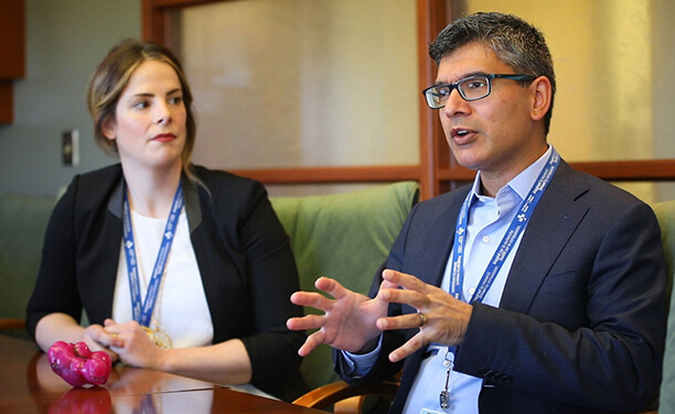
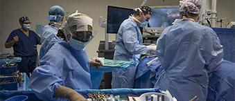
“This model helped to provide a good visual aspect. To have a model in my hands during surgery was incredible,” said Dr. Singh. “At the same time, we also had 3D images that I could look at on a TV screen in the operating room. It seems very futuristic, but in the operating room I was able to turn the image of the uterus at any angle or degree that I wanted, so I could see it from different perspectives, which helped during surgery.”

A picture might be worth a thousand words, but a 3D version is worth a million words. The 3D-printed models are not only helping surgeons, but also helping patients like Maureen understand their illness and prepare for their surgery. For patients, seeing a 3D model of the problem inside their bodies makes it tangible and real.

“Just before my surgery, Dr. Singh brought the model to me,” said Maureen. “He explained how he could use it in the surgery to see where the fibroids are, and he asked my permission to use it during the operation.”

She agreed, knowing that it would help other women suffering similar experiences. Dr. Singh successfully removed the fibroids, sparing Maureen from having a hysterectomy.

“We wanted to save her uterus in hopes that she can carry a pregnancy in the future, which wasn’t a hope for her up until this point,” said Dr. Singh.

“By working together with the 3D Printing Lab at The Ottawa Hospital, we’re going to be one of the first hospitals internationally to study how we can provide this improved care by using 3D-printed models in planning surgery for women’s health,” said Dr. Flaxman.

Dr. Sheikh said that, since the success of this first use of a 3D-printed model for gynecological surgery, the 3D Printing Lab is already working on a couple of other similar projects with the Minimally Invasive Gynecology team to offer other women alternatives to major surgery in the future.

Maureen was so grateful the gynecology team was able to spare her uterus, that she donated to the Gratitude Award Program to thank them.


The Ottawa Hospital is a leading academic health, research, and learning hospital proudly affiliated with the University of Ottawa.

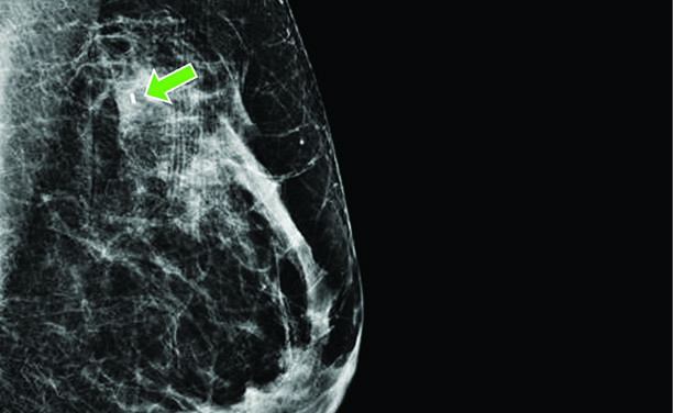
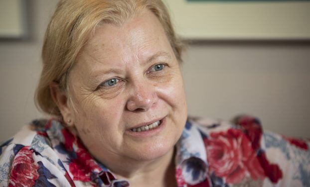
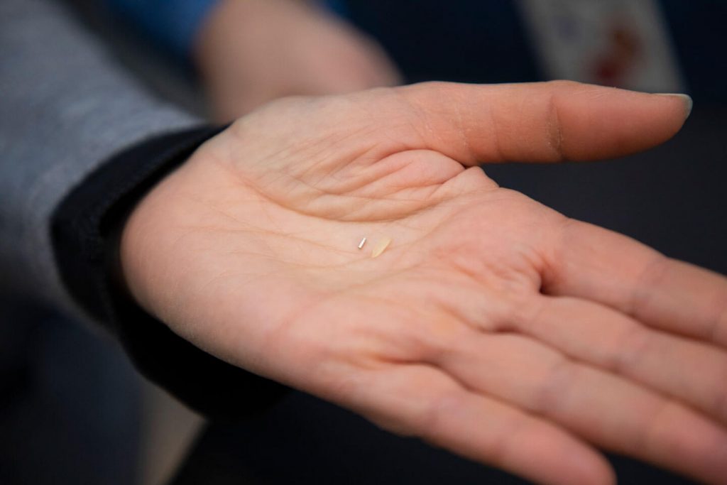
When a routine mammogram identified a small tumour, Rita Nattkemper was given an innovative option to mark its location for the surgery. A radioactive seed, the size of a pinhead, was injected directly into the tumour in her breast.

“All I have to say is it’s a painless procedure to get this radioactive seed in and it helps the doctor with accuracy,” said Rita. “And as he removes the mass, he’ll be removing the seed at the same time.”

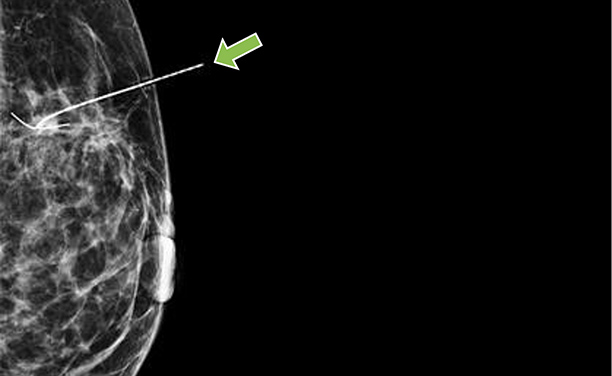
For the last 20 years, when a woman had a breast cancer tumour that was too small to feel or be seen in surgery (called a non-palpable tumour), she had to have a wire (known as a harpoon) implanted at the tumour site to locate it for the surgeon. The wire, which stuck out of the woman’s breast, had to be inserted the morning of her surgery. Then, women had to wait uncomfortably for surgery with the wire sticking out of their breast. To add to the discomfort, many women had to fast overnight to prepare for surgery, causing many to faint in the radiology suite at the sight of the wire protruding from their breast.

The procedure also posed other problems.

“Sometimes the wire moved. And sometimes, because of the location of the tumour, the wire might overshoot or undershoot the tumour, so ultimately there was an enormous amount of guess work involved in taking out a breast cancer tumour properly,” said Dr. Carolyn Nessim, surgical oncologist, and clinician-investigator in the Cancer Therapeutics Program at The Ottawa Hospital.

Dr. Nessim, and other breast oncology surgeons, wanted to find a better option.

That was where radioactive seeds came in. Radioactive seeds have been used for many years to treat prostate cancer. Multiple seeds are implanted in the prostate, where they emit radiation and kill the cancer. Then a procedure was developed for breast cancer patients using a radioactive seed to mark the exact location of small breast-cancer tumours. Using a mammogram for guidance, a radiologist places one seed, so tiny it can be safely injected with a needle, inside the tumour. It emits a very small amount of radiation that is picked up in the operating room with a small, handheld Geiger counter. After the piece of breast tissue with the radioactive seed is removed, the seed is separated from the tissue and appropriately disposed of, with every seed being accounted for.

Realizing the benefits of this procedure, The Ottawa Hospital began a radioactive seed program in 2015. One of the main benefits is that the seed can be placed up to a week before surgery, which makes the day of the operation easier for patients. A woman doesn’t have to wait for surgery with a wire sticking out her breast. From a logistical point of view, it’s easier to organize the procedure days in advance, and means more efficiency in the operating room. Dr. Nessim led a research study comparing seeds to wires, which showed the benefit of seeds.

“The results of the radioactive seed program have been uniformly excellent,” said Dr. Erin Cordeiro, breast surgical oncologist and senior clinician-investigator at The Ottawa Hospital. “We did a study that found that radioactive seeds were more cost effective and decreased wait times for patients on the day of surgery when compared to wires. And the patient experience has also been wonderful. Patients are very supportive of this.”

Rita agrees. She said the surgeon and radiologist both explained the procedure and put her at ease about it.

“I felt a minor pinch, and that was all I felt,” Rita said immediately after the procedure. “And the radiologist had the screen turned, so I could see where she put in the needle and left the seed. It was very easy, very quick, and very painless.”

Dr. Cordeiro said women are often concerned about the seed’s radioactivity, but the staff reassures patients the procedure is completely safe. The amount of radiation that is emitted in the week the patient has the implanted seed is less than having two mammograms.

“A woman can continue to hug her children and do everything in life she would normally do,” said Dr. Cordeiro. “No concerns from that point of view. It’s an extremely safe procedure. The vast majority of women have no concerns.”

Over the past year, 355 radioactive seed procedures have been performed at The Ottawa Hospital. Only two patients have refused the seeds and opted for the traditional wire instead.

Because of the program’s radioactive element, there were stringent guidelines around starting the program.

A multidisciplinary team of nuclear medicine, radiation safety experts, radiologists, pathologists, surgeons, technicians, and nurses were involved.

Key members of the team, led by Dr. Nessim, went to the Mayo Clinic in Rochester, N.Y., to learn how to implement the program. They then ran 15 training sessions for staff at The Ottawa Hospital. The radioactive seed program now “runs like a well-oiled machine,” said Dr. Nessim.

The Ottawa Hospital was the third centre in Canada to have a radioactive seed program, and is a leader in the procedure. Other health centres across the country are now adopting it and looking to The Ottawa Hospital for guidance in successfully implementing their program.


The Ottawa Hospital is a leading academic health, research, and learning hospital proudly affiliated with the University of Ottawa.
Creating
A Better
Tomorrow

Your support today unleashes
the potential of tomorrow

Every day people come to The Ottawa Hospital searching for answers; and every day, our innovative research brings hope to patients and their families. Every life changed, and every life saved through compassionate care and groundbreaking work at our hospital is made possible because of you.

We want you to be at our side; to help us push new discoveries and treatments forward, equip our team with the latest technology and equipment and ensure that our patients receive the very best care.

Creating Tomorrow

A Canadian-first clinical trial gives lymphoma patient a third chance
Faced with the return of his lymphoma for the third time in ten years, Owen Snider was running out of options. But there was new hope when he was accepted into a made-in-Canada CAR-T therapy clinical trial.
Catastrophic injuries require novel approach by our plastic surgery team
Karen Toop was hit by a snowplow while crossing the street in January 2012. She was critically injured when she arrived at our Trauma Centre. A multi-disciplinary team was ready to care for the injuries that some only see once in their career.
A meningioma tumour leaves mother facing blindness
With vision in her left eye deteriorating quickly, Michele Juma travelled from her home in Sault Ste. Marie to The Ottawa Hospital where she received specialized care after learning she had a meningioma tumour – and time was not on her side to save her vision.
From leukemia patient to multi-marathoner – with a walker
Leukemia patient, Bob Hardy, had a 40% chance of survival. But hope was restored after treatment at The Ottawa Hospital.
Celebrating a “re-birthday” each year since having a cancerous brain tumour removed
Ten years ago, The Ottawa Hospital saved Kimberly Mountain’s life after the discovery of cancerous brain tumour. Today, she’s confident if the cancer comes back, The Ottawa Hospital will be ready to save her again.
Hope despite aggressive skin cancer diagnosis
Diagnosed with a stage 4 melanoma at the age of 62, Dan Collins feared for his life when he learned about the aggressive form of cancer. However, immunotherapy treatment gave him a reason to hold out hope.

Important Notice - Canada Post operations have resumed, but mail delivery may still be affected. For more details on online and in-person donation options, please click here.